Oxfordian
Hatching
- Feb 22, 2017
- 2
- 0
- 7
I periodically lose a hen to "messy tail feather syndrome". This happens to my hens who are over 3 years old. Rather than episodic diarrhea, the messy tail feathers return even when the hen has been bathed and is clean. These hens have a slow decline resulting in an enlarged and very firm abdomen, lowered tail, little to no appetite, darkened comb. It has happened to hens of every breed I've had; I have about 1 hen a year that travels this road. My very favorite hen (Golden Comet) started the slow decline. I took her to the vet (Auburn Animal Hospital in MA) and we treated her for a month with 2.4 ml Sulfatrim twice a day and .75 ml metcam once a day. She improved when on the full dose of the meds but as soon as I backed off the Sulfatrim she declined rapidly. She also started to really hate the oral meds and I had a hard time getting them in her with a syringe or mixing them into a mash. It was clear she was not going to recover and I decided to put her down and do an autopsy. I've attached the notes and resulting photos. My main question was: was she suffering one incident of egg yolk peritonitis which created a well-established infection or was she having multiple episodes. The answer was clearly that she had many egg yolks in her peritoneal cavity and nothing short of a hysterectomy was going to stop the mis-firing of yolks. I've concluded that today's breeds are bred for productivity and with age come reproductive problems, especially for the good layers. Very sad end for these wonderful hens. Below are the notes from the autopsy:
Masked/maintained iso/02. Pentobarbital 2 ml IV basilar vein R wing. Removed 190 ml yellow turbid fluid with fibrin strands via coelomocentesis. Opened coelom; air sacs, liver, spleen, kidneys NSF. Ventriculus NSF, SI loops with some firm white nodules dispersed over surface and throughout mesentery. Large volume (approx 250 ml) yellow fluid with multiple large fragments of yellow friable material, and 3 free larger, more clearly identifiable as yolk structures. Ovary with multiple dark, uniform sized follicles, as well as 2 attached larger yolks. Oviduct very thickened, firm, and also covered in white, firm nodules. Wall of oviduct very thickened, mild caseous yellow material within. Clear fluid with granular material in crop. RIO bacterial salpingitis with secondary EYP (multiple free yolks). Clinical improvement on antibiotics may have been secondary to salpingitis resolution and possible immunomodulation by the sulfa drug component. Cannot rule out viral (esp Marek's) involvement, although no other gross necropsy findings were seen to indicate Marek's (ie enlarged sciatic nn or other peripheral nn; iridal changes, or primary GI lesions) 2/20/17
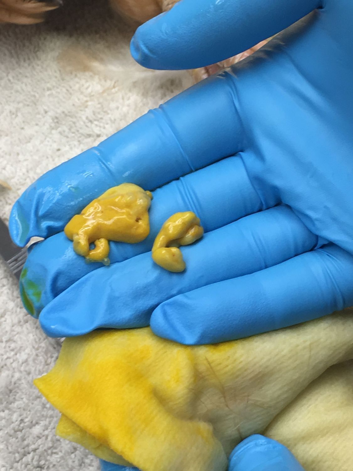
Egg yolk from coelom
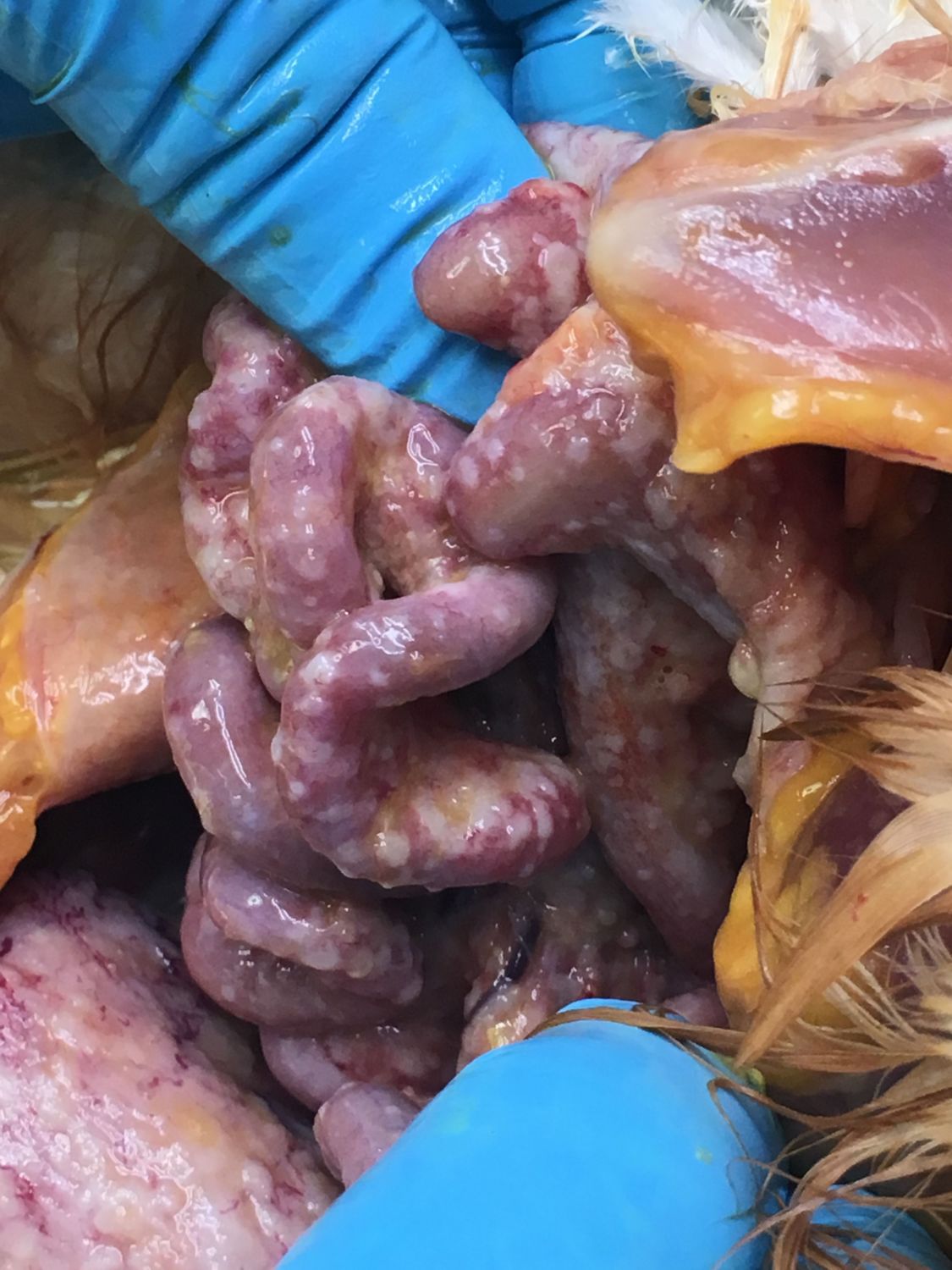
Inflamed oviduct and free yolk

Normal air sac

Normal liver

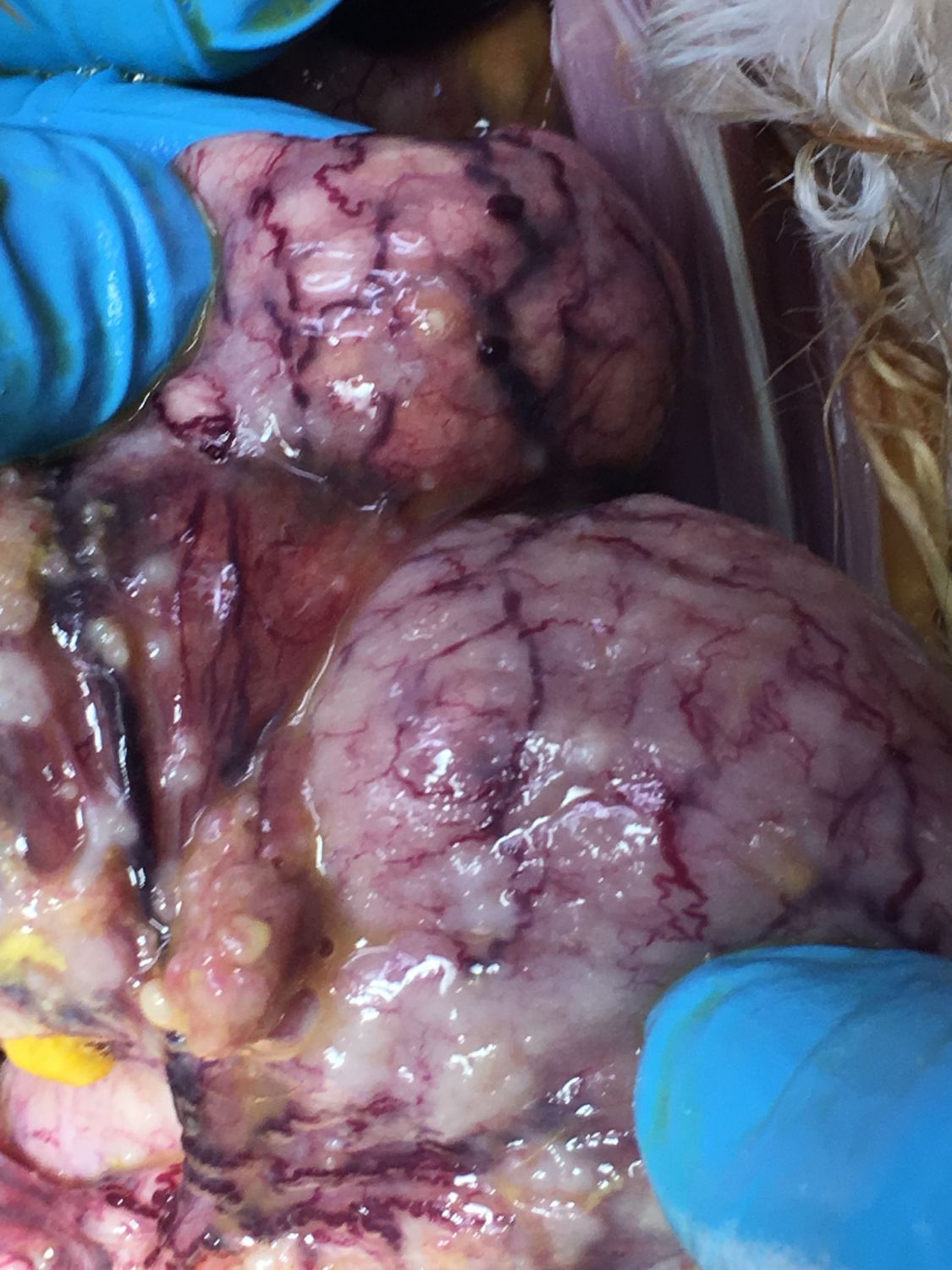
Ovary and oviduct

Ovary buried in fluid and free yolk

Oviduct impacted area
Oviduct
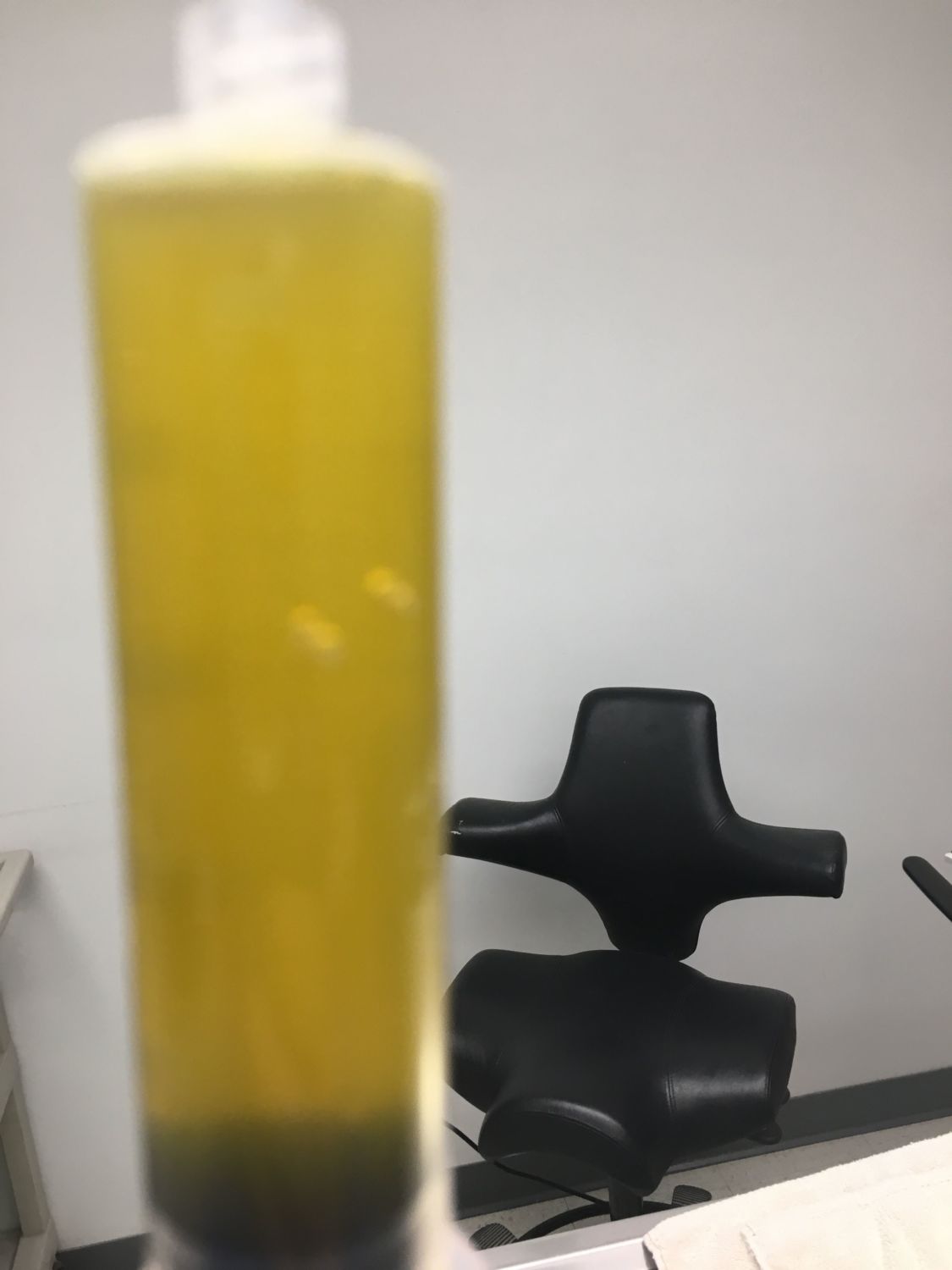
Peritoneal fluid - .5 liter was removed
Masked/maintained iso/02. Pentobarbital 2 ml IV basilar vein R wing. Removed 190 ml yellow turbid fluid with fibrin strands via coelomocentesis. Opened coelom; air sacs, liver, spleen, kidneys NSF. Ventriculus NSF, SI loops with some firm white nodules dispersed over surface and throughout mesentery. Large volume (approx 250 ml) yellow fluid with multiple large fragments of yellow friable material, and 3 free larger, more clearly identifiable as yolk structures. Ovary with multiple dark, uniform sized follicles, as well as 2 attached larger yolks. Oviduct very thickened, firm, and also covered in white, firm nodules. Wall of oviduct very thickened, mild caseous yellow material within. Clear fluid with granular material in crop. RIO bacterial salpingitis with secondary EYP (multiple free yolks). Clinical improvement on antibiotics may have been secondary to salpingitis resolution and possible immunomodulation by the sulfa drug component. Cannot rule out viral (esp Marek's) involvement, although no other gross necropsy findings were seen to indicate Marek's (ie enlarged sciatic nn or other peripheral nn; iridal changes, or primary GI lesions) 2/20/17
Egg yolk from coelom
Inflamed oviduct and free yolk
Normal air sac
Normal liver
Ovary and oviduct
Ovary buried in fluid and free yolk
Oviduct impacted area
Oviduct
Peritoneal fluid - .5 liter was removed


